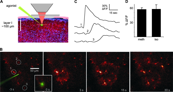
Figure 1.
Noradrenergic agonists elicit Ca2+ waves in cortical astrocytes. (A) The image at left illustrates the experimental setup with a cranial window over the somatosensory cortex and a glass electrode containing agonist positioned in the molecular layer (50–80 μm deep). (B) Series of images demonstrating a Ca2+ wave to pressure ejection of 200 μM methoxamine. (C) Changes in Ca2+ over time corresponding to numbered cells circled in (B) showing the time course of wave propagation. (D) Histogram showing that both α- (34 cells in 5 animals) and β-agonists (200 μM; 27 cells in 4 animals) are capable of eliciting Ca2+ responses in cortical astrocytes. meth, methoxamine; iso, isoproterenol.

