Figure 2.
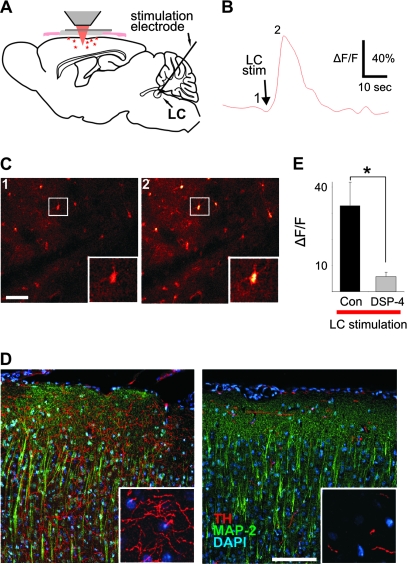
Direct LC stimulation elicits Ca2+ transients in cortical astrocytes. (A) Schematic illustrating experimental setup. (B) Average fluorescence of the image field in (C) over time showing Ca2+ response to LC stimulation. (C) Fluo-4 labeled astrocytes taken at time points indicated by numbers in (B). Insets are blown-up view of boxed area in pictures. Scale bar 50 μm. (D) Images from coronal sections through somatosensory cortex illustrating the effect of DSP-4 on LC projection neurons as labeled by antibodies to TH and MAP-2 with DAPI labeling of nuclei for orientation. Scale bar 100 μm (20 μm inset). (E) Consistent with the significant reduction in LC projection neurons and despite almost twice the stimulation intensity (216 ± 33 vs. 400 ± 39 μA; P = 0.004, t-test), there is a significant reduction in the cortical Ca2+ response to LC stimulation in DSP-4 treated animals (P = 0.013, t-test).

