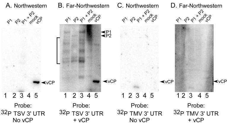
Figure 3.
RNA-RdRp interactions detected by far-northwestern blotting are specific to AMV and ilarvirus RNAs. P1, P2, P1/P2, mock extract, and AMV virion coat proteins were separated by SDS-polyacrylamide gel electrophoresis, transferred to nitrocellulose membrane, and successively denatured and renatured prior to probing with radiolabeled RNA only (panels A and C) or radiolabeled RNA plus soluble AMV coat protein (panels B and D). The probes used for the experiments were the 3' untranslated region of tobacco streak virus (TSV) RNA 4 (panels A and B) or the 3' untranslated region of tobacco mosaic virus RNA (panels C and D). The bracket indicates labeled RNA associated with protein bands that are likely to be hydrolysis products of the P1 and P2 proteins, which were found to be relatively unstable on storage.

