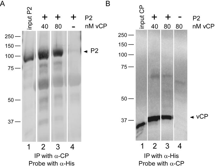
Figure 5.
Analysis of AMV coat protein-polymerase interactions by co-immunoprecipitation. AMV coat protein (40 nM or 80 nM) was added to soluble P2 protein. Complexes were immunoprecipitated with anti-AMV coat protein or anti-6his antibodies. The precipitates were collected, separated by SDS-PAGE, transferred to nitrocellulose membrane, and probed. A) Immunoprecipitation with anti-coat protein antibody, followed by electrophoresis, transfer, and probing with anti-6his antibody to detect P1 and P2 proteins. Lane 1: P2 protein only (input); lanes 2 and 3: P2 protein with 40 nM or 80 nM virion coat protein added, respectively; lane 4: P2 protein extract without added coat protein. B) Immunoprecipitation with anti-6his antibody, followed by electrophoresis, transfer, and probing with anti-coat protein antibody. Lane 1: Virion coat protein only; lane 2: 40 nM virion coat protein with P2 protein; lane 3: 80 nM virion coat protein plus P2 protein; lane 4: 40 nM virion coat protein, without added P2 protein. There was some distortion (“frowning”) of the gel shown in Figure 5B; therefore, the bands in lane 4 do not align perfectly with the other lanes. However, the results show that there is little detectable coat protein signal present when P2 protein was omitted from the immunoprecipitation reaction.

