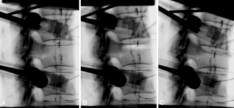
Fig. 2.
Digital fluoroscopy images of a specimen. a Intact specimen. The bilateral loading cables, coated with radiopaque barium solution, are visible on the X-ray images. b Radiographic appearance of the void created under the upper endplate of the middle vertebra. c Image of the wedge fracture affecting only the upper part of the index vertebra

