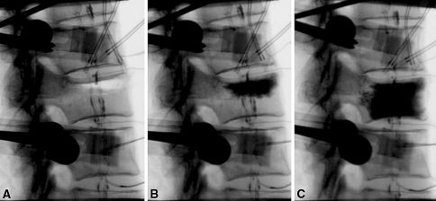
Fig. 3.
Digital fluoroscopy images of a specimen a Reduction of anterior wall height and vertebral kyphosis angle with extension of the specimen while under 150 N preload. b Cement augmentation of the fracture. c Image showing the uniform distribution of cement under both endplates after careful abrasion of the un-fractured endplate

