Fig. 1.
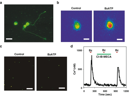
The P2X7 receptor on rat retinal ganglion cells. a Immunohistochemical staining for the P2X7 receptor on rat retinal ganglion cells isolated from the retina at postnatal day (PD) 8 and grown for 2 days on a coverslip treated with poly-L-lysine and laminin. Three cell bodies are in view; neurites projecting from two cells also stained for the P2X7 receptor. Of note, the growth cones on the neurites stained brightly. Scale bar = 20 µm. Background staining produced by reaction with the coverslip coating is seen. b Pseudocolor image of a rat retinal ganglion cell loaded with the calcium indicator fura-2 and excited at 340 nm. Application of 50 µM BzATP increased the signal from the cell (with red indicating a greater intensity), indicative of an increase in intracellular calcium levels. Bar = 10 µm. c Incubation with 50 µM BzATP decreased the number of viable ganglion cells. Ganglion cells were labeled in vivo by injection of fluorogold derivative aminostilbamidine into the superior colliculus followed by retrograde transport of the dye into the ganglion cell bodies, ensuring identification of ganglion cells when dissociated retinal cells were exposed to drugs in vitro. The number of fluorescent ganglion cells remaining after 24 h in the presence of BzATP in vitro was significantly less than in control. Bar = 100 µm. d The A3 adenosine receptor attenuated the calcium rise triggered by stimulation of the P2X7 receptor. A brief application of 50 µM BzATP led to a rapid and reversible rise in calcium within a rat ganglion cell, as calibrated from the ratio of fluorescence excited at 340–380 nm in a fura-2-loaded cell. The response to BzATP was prevented by 1 µM of the A3 adenosine receptor antagonist Cl-IB-MECA, but was restored upon removal of Cl-IB-MECA

