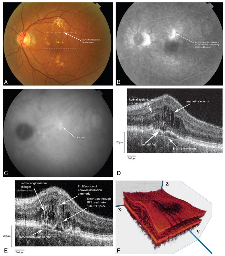
Fig. 4.
Patient 2. A, Color fundus photograph of retinal angiomatous proliferation (RAP) shows intraretinal hemorrhages overlying a grayish mound of deep angiomatous changes just superior to the fovea. The dotted lines correspond to the Fourier-domain optical coherence tomography (Fd-OCT) images presented. B, Fluorescein angiogram shows intense hyperfluorescence of the RAP lesion with ill-defined zone of leakage around the angiomatous changes. C, Indocyanine green angiogram shows a focal area of intense hyperfluorescence (“hot spot”). D, Fd-OCT image (small dashed line) shows multifocal areas of high reflectivity corresponding to the angiomatous changes. Adjacent to the RAP lesions are areas of intraretinal edema. There is tenting up of the retinal pigment epithelium (RPE) under the neovascular proliferation, forming a pocket of subretinal fluid. Note that Bruch’s membrane is intact. E, Fd-OCT B-scan (large dashed line) shows the extension from the intraretinal neovascularization through a break in the RPE to an early lesion in the sub-RPE space. Note that Bruch’s membrane is still intact. F, Three-dimensional reconstruction of the macula with cut through the RAP complex. The B-scans are taken from the x plane.

