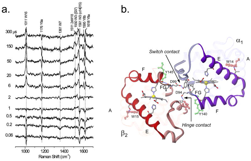
Figure 5.
a) Pump-probe 229 nm UVRR difference spectra of the HbCO photoproduct at the indicated time delays, showing the evolution of Tyr and Trp signals. b) Ribbon diagram of the α1 and β2 subunits in deoxyHb, showing Trpβ37 and Tyrα42 quaternary H-bonds at the hinge and switch contacts. These are formed in sequence, after initial deligation-induced rotation of the EF helix ‘clamshells’, breaking A…E and F…G interhelical H-bonds (involving W14 and Y140 in the α subunit, and W15 and Y145 in the β subunit [30].)

