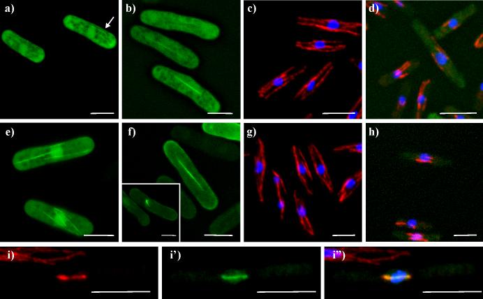
Figure 3. Klp6-motor-EGFP and Klp6-motor-neck-EGFP shorten microtubules.
a, b) live cells expressing Klp6-motor-EGFP. a) Interphase cells; arrow indicates cell where some fluorescence is concentrated on fibers that resemble MTs; b) mitotic cells. c, d) fixed cells stained for tubulin (red), and DNA (blue); c) cell with Klp6-motor-EGFP, repressed by the addition of thiamine; d) cell expressing Klp6-motor-EGFP. e,f) live cells expressing Klp6-motor-neck-EGFP; e) interphase cells; f) mitotic cells, inset; cell with a monopolar spindle. g-i) fixed cells with Klp6-motor-neck-EGFP (green), tubulin staining (red), and DNA staining (blue); g and h display results from same experiment as c and d. i) tubulin (Texas Red) channel; i') Klp6-motor-neck-EGFP (FITC) channel; i”) combined channels with DNA (DAPI) DNA. White bars = 5μm.

