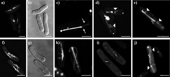
Figure 4. Localization of Klp5-full-length-EGFP and Klp6-full-length-EGFP.
a-e) Klp5-full-length-EGFP imaged in live cells. a) cells with one or two large bundles of MTs; b) bright field image of cells in first panel; c) cell with single large bundle of MTs, with individual MTs splaying out from the bundle (arrows); d) cells stained for DNA (Hoechst) show that bundles are not spindles; e) mitotic cell in late anaphase B. f-j) Klp6-full-length-EGFP imaged in live cells. f) two cells with shorter-than-normal peri-nuclear MTs and dots; b) bright field image of previous panel; h) cells with MT bundles and long, curled MTs; i) two cells with short spindles; j) cell stained for DNA (Hoechst) with a somewhat abnormal, late mitotic spindle. White bar = 5μm.

