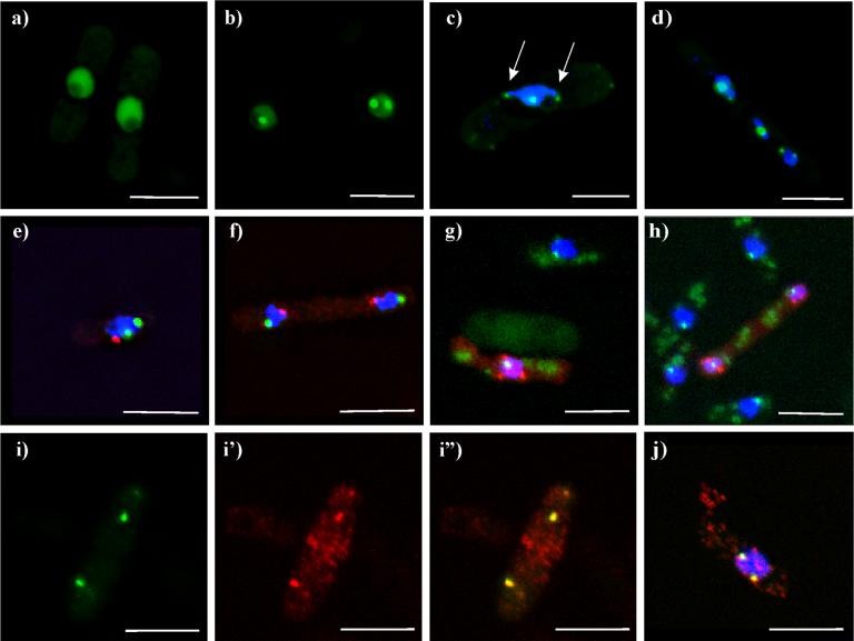
Figure 5. Klp5-tail-EGFP localization to telomeres.
a-d) live cells expressing Klp5-tail-EGFP. a) cells with nuclear staining, excluding the nucleolus; b) cell in late telophase with nuclear staining and dots in each nucleus; c-h,j) cells stained with Hoechst 33342 for DNA (blue); e-f) simultaneous staining with the spindle pole body marker, Sad1p (red). 5c shows nuclear dots associated with DNA protrusions (arrows); d) klpΔ cell with DNA staining, showing GFP dots associated with each chromosome. e) interphase cell; f) late mitotic cell. g, h) Klp5-tail-Pk1 (red) with the kinetochore marker Mis12-GFP (green), and DNA (blue). 5g and h) interphase cell and late mitotic cell, respectively, both with Klp5-tail-Pk1 stained red. i-j) Simultaneous staining of Klp5-tail-Pk1 (red) with the telomere marker Taz1-GFP (green). i) interphase cells, Taz1-GFP; i') Klp5-tail-Pk; i”) combined channels. j) mitotic cell, combined channels. White bar = 5μm.

