
Fig. 3.
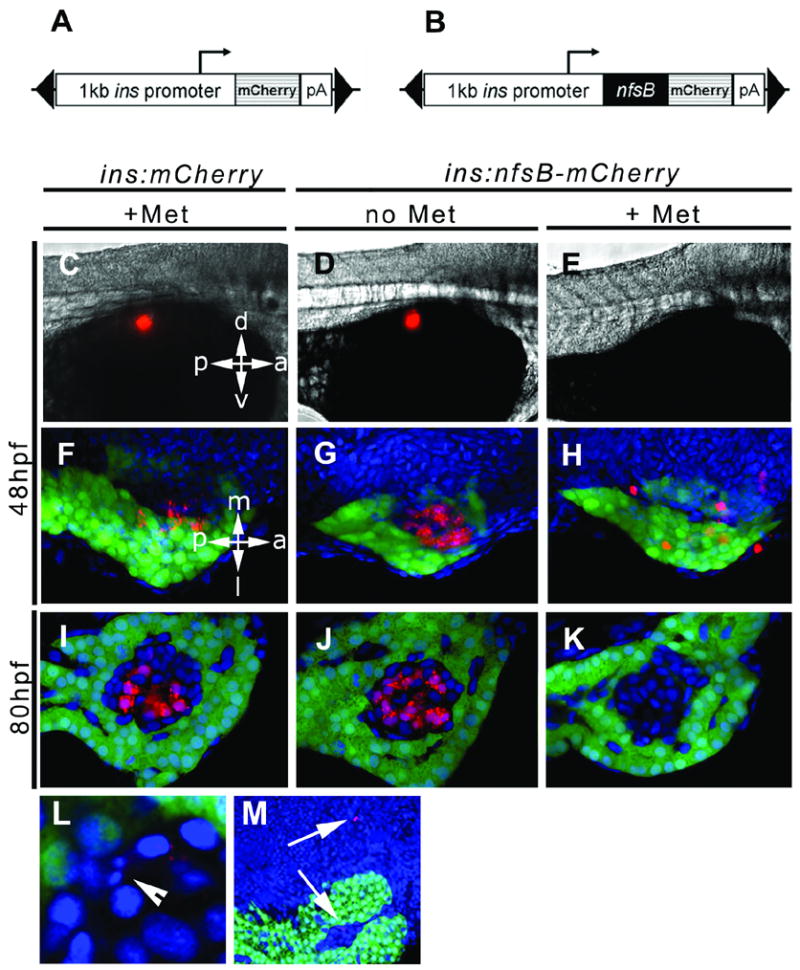
The ins:nsfB-mCherry transgene leads to prodrug dependent loss of β cells. Schematic of constructs used to drive β cell expression of mCherry in (A) and the fusion protein NTR-mCherry in (B). The expression cassettes were cloned inside of a Tol2 transposable element (black triangles). In the experiment shown here, two groups of controls were used, ins:mCherry embryos incubated in Met (+Met, C, F, I) and ins:nfsB-mCherry without prodrug (no Met, D, G, J). Experimental groups consisted of ins:nfsB-mCherry embryos incubated for 24 hrs in Met (+Met, E, H, K). Two separate time points were chosen for addition of prodrug as shown, incubation starting at 24hpf and finished by 48hpf (48hpf, C to H) and starting at 56hpf and finishing at 80hpf (80hpf, I to K). Embryos were photographed by either epi-fluorescence in (C) to (E), or by confocal microscopy on isolated pancreata plus adjacent endoderm in (F) to (M). Red fluorescence indicated presence of mCherry (C, F, I) or NTR-mCherry fusion protein (D, E, G, H, J, K, L and M). eGFP marks the exocrine pancreas with green fluorescence and nuclei are stained blue (Hoescht). (L) Example of fragmented nucleus indicative of apoptosis (arrowhead) and cell debris inside and outside of islet in (M) (arrows). Orientation as indicated, d = dorsal, v = ventral, a = anterior, p = posterior, m = medial and l = lateral.