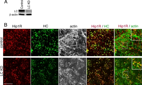
FIGURE 7.
Immunofluorescence of Hip1R, actin, and clathrin following cellular depletion of clathrin light chains. HeLa cells were transfected with siRNAs targeting both clathrin light chain a (LCa) and clathrin light chain b (LCb) to knock down all clathrin light chains (LC KD) or with control (scrambled) siRNA at the same concentration and cultured for 72 h. A, HeLa cells were lysed after siRNA treatment, and protein levels were determined by immunoblotting. The indicated proteins were detected by a rabbit polyclonal antiserum against a conserved sequence shared by LCa and LCb (LC) (29) and β-actin (Sigma). Actin is shown as a loading control. B, cells treated with siRNA against clathrin light chain (LC KD) or with control siRNA were labeled for immunofluorescence with antibodies against clathrin heavy chain (HC) (green) and Hip1R (red). Actin was detected with fluorescent phalloidin and is shown in black and white images in the center panels or in green in the far right panels. Merged images of the indicated proteins are shown in the two right-hand panels of each row, with yellow indicating overlap of red and green labeling. In the far right panels, the central boxed area is magnified in the upper righthand corner. Bar, 10 μm.

