FIGURE 2.
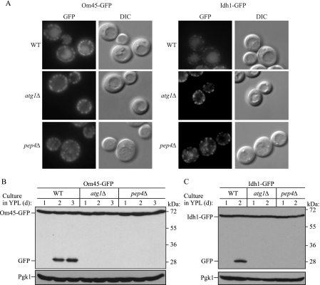
Monitoring mitophagy using C-terminal GFP-tagged mitochondrial protein processing at post-log phase. Wild-type (WT), atg1Δ, and pep4Δ strains expressing Om45-GFP or Idh1-GFP were cultured in YPL medium for 1–3 days. The localization of GFP was visualized by fluorescence microscopy at day 2 (A), and GFP processing was monitored by immunoblotting with anti-GFP and anti-Pgk1 (loading control) antibody and antiserum, respectively, at the indicated days in the strains expressing Om45-GFP (B) or Idh1-GFP (C). DIC, differential interference contrast.

