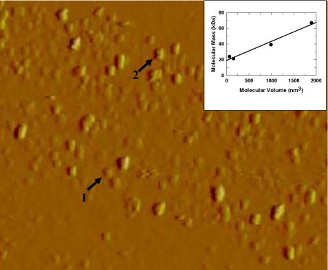
FIGURE 5.
AFM images of human wild-type GST π (from a solution of 0. 25 mg/ml). Image size 25 nm × 4.5 μm. Arrow 1: monomer; arrow 2: dimer. Inset: the calculated volume of proteins imaged by AFM plotted against the molecular mass. The proteins used are xylanase from T. longibraciatum (21.6 kDa), polygalacturonase from A. nigar (24.0 kDa), xylanase from C. japonicus (39.2 kDa), and bovine serum albumin (67.0 kDa).

