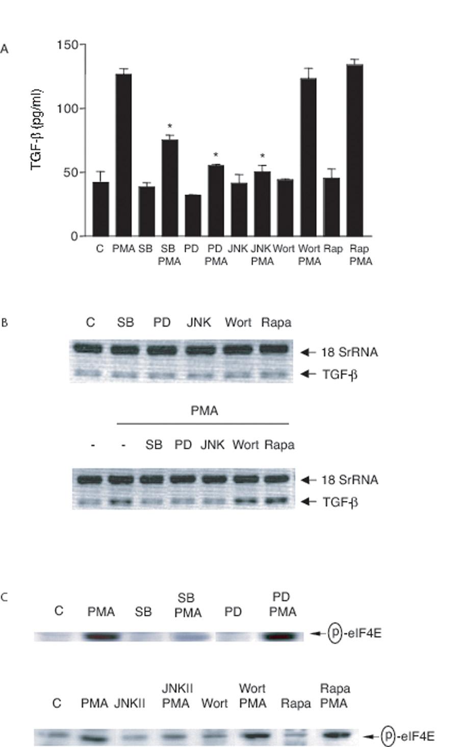
FIGURE 8. Effect of inhibitors on PMA-induced TGF-β production.
A, 3T3TβRII cells were pre-treated with rapamycin (Rapa, 100 nM) overnight, SB 203580 (SB, 10 µM), PD 98059 (PD, 30 µM), JNK inhibitor II (JNKII, 30 µM) or wortmannin (Wort, 100 nM) for 1 h and then stimulated with PMA (100 nM) for 18 h. Total TGF-β in the conditioned medium was analyzed by ELISA. *, significantly different from PMA alone. B, 3T3TβRII cells were pre-treated with rapamycin (Rapa, 100 nM) overnight, SB 203580 (SB, 10 µM), PD 98059 (PD, 50 µM), JNK inhibitor II (JNKII, 50 µM) or wortmannin (Wort, 100 nM) for 1 h and then stimulated with PMA (100 nM) for 4 h. TGF-β mRNA levels were analyzed using Relative Quantitative RT-PCR. C, 3T3TβRII cells were pre-treated with rapamycin (Rapa, 100 nM) overnight, SB 203580 (SB, 10 µM), PD 98059 (PD, 50 µM), JNK inhibitor II (JNKII, 50 µM) or wortmannin (Wort, 100 nM) for 1 h and then stimulated with PMA (100 nM) for 30 min. Total cell lysates were immunoblotted for phospho-eIF4E.

