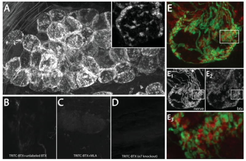
Figure 2.

BTX specifically labels postsynaptic AChR α7. (A) BTX labeling in the SMG. 10 nM TRITC-BTX was added to the neck of an anesthetized wild-type mouse. After BTX labeling (1 h), postganglionic neurons display punctate fluorescent labeling. Inset shows clusters of fluorescence on the surface of two postganglionic neurons. (B) Pre- and coincubation with a saturating dose of unlabeled BTX blocks TRITC-BTX labeling. (C) Methyllycaconitine (MLA), a reagent known to specifically block the interaction between BTX and AChR α7, blocks TRITC-BTX labeling in SMG. (D) BTX labeling is not present in the SMG of genetically modified animals (red) lacking the AChR α7 subunit. Image acquisition parameters were the same for (A) through (D). (E) BTX labeling (red) is selectively opposed to presynaptic terminals (green). (E1,2) Single channel images from (E) depicting YFP labeled preganglionic axons (E1) and BTX labeled postsynaptic AChRs (E2). (E3) Single optical section of the boxed region in (E) shows that YFP-labeled presynaptic terminals are closely juxtaposed to BTX labeled regions.
