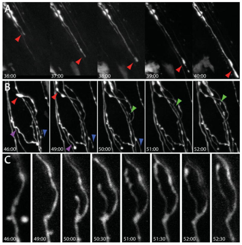
Figure 4.

In vivo time-lapse imaging of axon growth and synapse formation. Preganglionic axons were crushed with a blunt forceps along the salivary ducts at time 0:00 and imaged at the indicated times after crush (hr:min). (A) 1 [1/2] days later, regrowth could be observed as preganglionic axons grew toward postganglionic neurons. Red arrows indicate sites of axon advance. (B) Axons rapidly reinnervate postsynaptic postganglionic neurons (dark region in the middle of the image) by sending processes that wrap around the soma. Colored arrows indicate sites of axon elongation. (C) When preganglionic axons contact postganglionic neurons, presynaptic boutons appear along axonal branches as axons change from a uniform to a scalloped shape.
