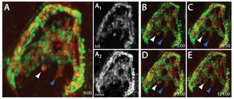
Figure 5.

In vivo time-lapse imaging of pre- and postsynaptic structures. YFP-labeled presynaptic axons and BTX-labeled AChRs were imaged over time. 647-BTX was added to the neck for 1 h, and then washed out with Ringer's solution. YFP-labeled preganglionic axons (A2) and 647-BTX-labeled postsynaptic AChRs (A1) were simultaneously imaged on a confocal microscope. (B–E) After the first image, the wound was sutured closed and the animal was allowed to recover. Synapses were relabeled with BTX for each successive imaging session. Arrows indicate individual synaptic regions that could be identified for several days. The white arrow indicates stable presynaptic terminals and postsynaptic AChRs, while the blue arrow shows a region where presynaptic terminals are dynamic but postsynaptic AChRs are stable. Times correspond to hr:min.
