
Table 1.
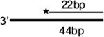
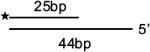
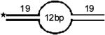
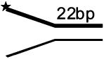
Structures of DNA Substrates for Helicase Assays
| Name | Structure | Name | Structure |
|---|---|---|---|
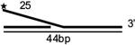
| 3′-overhang (1+2) |
 |
4bp bubble (10+11) |
|
| 5′-overhang (2+3) |
 |
12bp bubble (12+13) |
 |
| Blunt-end duplex (4+5) |
21bp bubble (14+15) |
||
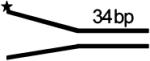
| forked duplex (6+7) |
 |
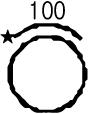
100bp M13mp 18 Partial duplex |
 |
| forked duplex (8+9) |
 |
FLAP (2+16+18) |
|
| FLAP (2+16+17) |
 |
Holliday-Junction (19+20+21+22) |
 |
| Synthetic Replication fork (2+14+15+16) |
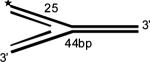 |
★
Radioactive label at 5′