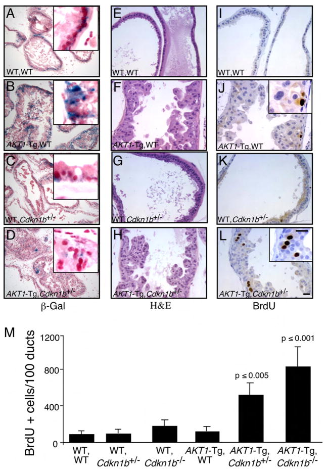
Figure 2. Genetic inactivation of Cdkn1b rescues cell from senescence and increases proliferation in AKT1-Tg mice.
(A–L) Twelve hours after BrdU administration, mice were sacrificed and the VPs of wild-type (A, E, I), AKT1-Tg (B, F, J), Cdkn1b+/− (C, G, E) and AKT1-Tg, Cdkn1b+/− (D, H, L) mice were stained with either β-Gal (A– D), H&E (E–H) or with anti-BrdU antibody (I–L). Scale bar, 50 μM; insert 100μM.
(M) The numbers of BrdU stained cells per 100 ducts was determined by manual counting of BrdU positive cells in all lobes of VP. Data are presented as mean ± SD.

