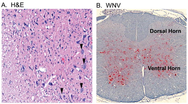
Figure 2.
Coronal section of spinal cord at L2–L4 vertebrae from a hamster at the first day of paralysis (8 d.p.i.) challenged by injection of WNV (101.8 p.f.u.) in the cord at T8 verbetrae. (A) H&E staining on the first day of paralysis at 8 days after challenge. (B) Alkaline phosphatase detection of WNV env antigen (red) at 5 days after challenge.

