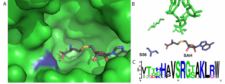
Fig. 6. Crystallographic and bioinformatics analysis of the flavivirus methyltransferase active site.
(A) Pocket of Dengue Virus methyltransferase, with SAH in atomic coloring and S56 in blue. Hydrogen bonds between SAH and S56 are shown by yellow dotted lines (B) Direct interactions between S56 (dark blue) and SAH in the active site of Dengue Virus methyltransferase. The atoms shown in green are the canonical KDKE active site of all methyltransferases. The orientation of (A) and (B) are the same. (C) Logo analysis of the region around S56 from 715 distinct flaviviral genomes. The phosphorylated serine (S56) is starred. (alignment: http://athena.bioc.uvic.ca/workbench.php?tool=vocs&db=, for Logo: http://weblogo.berkeley.edu/).

