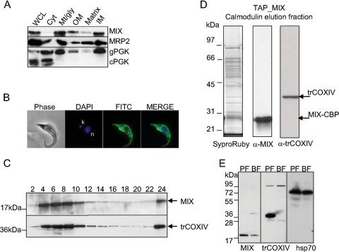
FIG. 6.
Expression, localization, and purification of the TAP-tagged MIX complex. (A) Subcellular fractionation and Western blotting using anti-MIX antibody indicates that MIX is localized to the mt inner membrane. The 25-kDa mt MRP2 protein and 56-kDa glycosomal phosphoglycerate kinase (gPGK) and 45-kDa cytosolic phosphoglycerate kinase (cPGK) isozymes were used as marker proteins. WCL, whole-cell lysate; Cyt, cytosolic fraction; Mt/gly, mt and glycosomal fraction; OM, outer membrane fraction; IM, inner membrane fraction. (B) Immunolocalization of tagged MIX protein shows an even distribution in the reticulated mitochondrion of procyclic T. brucei. Phase-contrast light microscopy shows procyclic T. brucei cells, DAPI staining is specific to DNA contents (n, nucleus; k, kinetoplast), and fluorescein isothiocyanate (FITC)-conjugated secondary antibody shows mt localization of the target protein. (C) Sedimentation analysis of MIX and the trCOXIV subunit of the cyt c oxidase complex. The cleared lysate of hypotonically purified mitochondria was loaded onto a 10 to 30% glycerol gradient and fractionated. The even fractions were screened for the presence of MIX and trCOXIV proteins using specific polyclonal antibodies against these proteins. (D) The TAP-tagged MIX complex was separated on a 10 to 14% polyacrylamide Tris-glycine gel and stained with Sypro Ruby. Positions of bait protein (MIX-CBP) and trCOXIV were determined by immunoblot analysis. The sizes of the protein markers are indicated on the left. (E) MIX is expressed throughout the Trypanosoma life cycle, with reduced expression in the BF. Western analysis using anti-MIX, anti-trCOXIV, and anti-Hsp70 as a loading control was carried out on lysates from the PF and BF.

