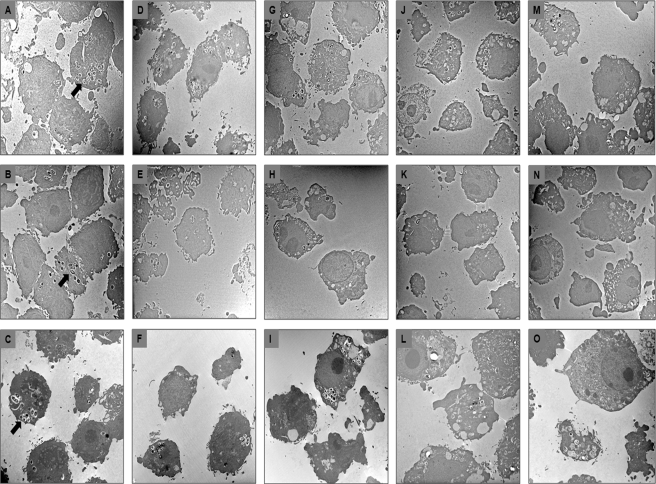
FIG. 5.
Transmission electron microscopy of RAW 264.7 macrophages at 0 h (A, B, and C), 2 h (D, E, and F), 24 h (G, H, and I), 48 h (J, K, and L), and 72 h (M, N, and O) after infection with E. faecalis strains E99 (A, D, G, J, and M), DBS01 (B, E, H, K, and N), and DBS01(pGT101) (C, F, I, L, and O). Arrows indicate bacterial cells inside macrophages.

