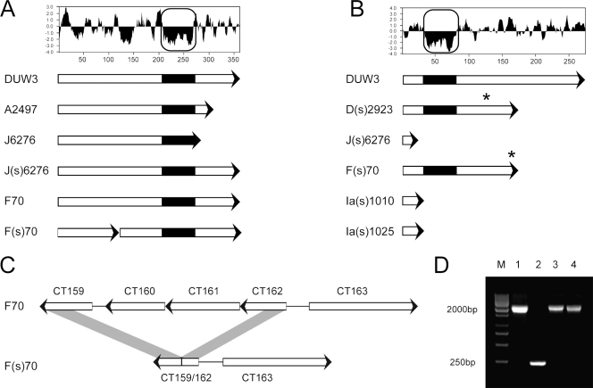
FIG. 2.
ORF analysis of regions showing variation found in the matched pair isolates. (A) Kyte-Doolittle hydrophilicity plot of gene CT135, and gene representations of lab strain D/UW3 (DUW3), ocular isolate A2496 (11), and matched pair isolates. (B) Kyte-Doolittle hydrophilicity plot of the predicted CT119 (incA) protein sequence, and ORF representations of lab strain D/UW3 and clinical isolates showing the IncA-negative phenotype. The boxed regions in panels A and B highlight a putative Inc motif. Asterisks indicate locations of frameshift mutations leading to downstream truncation. (C) A region of the plasticity zone that is deleted in isolate F(s)/70 relative to strain F/70, resulting in putative CT159/CT162 fusion. (D) PCR analysis confirming the deletion found in isolate F(s)/70, with the length of each product indicated to the left. Lanes: M, molecular size markers (M); 1, strain F/70; 2, strain F(s)/70; 3, strain F(s)/4022; 4, strain F(s)/8068.

