
FIG. 3.
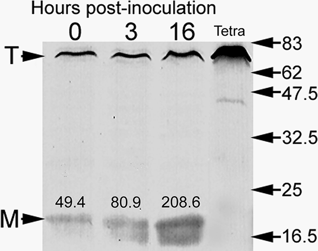
Immunoblot showing that expression of MagA (a protein marker of MIF development in Philadephia-1 strains) is upregulated during the interaction of Lp1-SVir with T. tropicalis. Samples were loaded based on CFU counts (5 × 10⁴/lane). The mean CFU/ml values of the two independent samples pooled at each time point were 6.3 ± 1.52 × 10⁵ (time zero), 8.5 ± 1.95 × 10⁵ (3 h), and 18.3 ± 6.00 × 10⁵ (16 h). The T arrowhead points to a Tetrahymena protein band recognized by the MagA polyclonal antibody. This protein band is well-labeled in samples containing ciliates only (lane labeled Tetra). The M arrowhead points at the position of MagA. Densitometry values in arbitrary IOD units are shown above each MagA band. The positions and molecular weights (×1,000) of broad-range, prestained protein standards are indicated on the right side.