FIG. 1.
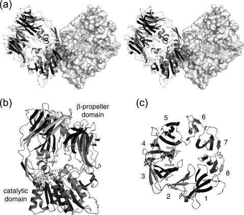
Crystal structure of Stenotrophomonas DPIV. (a) The dimer structure of Stenotrophomonas DPIV is shown by a stereo diagram. One of the two subunits is shown as a ribbon model, and the other is shown as a surface diagram. The diagram was drawn with the programs MOLSCRIPT (31) and Raster3D (40) and rendered with the program PYMOL (http://www.pymol.org/). (b) The subunit structure is shown as a ribbon model. The subunit was composed of two domains, the N-terminal β-propeller domain (top) and the C-terminal catalytic domain (bottom). The catalytic triad and two glutamate residues at the active site are shown as ball-and-stick models. (c) Ribbon model of the β-propeller domain, looking down upon the model as shown in diagram b, with assigned blade numbers. The last two diagrams were drawn with the program POVSCRIPT+ (15) and rendered with the program POVRAY (http://www.povray.org/).

