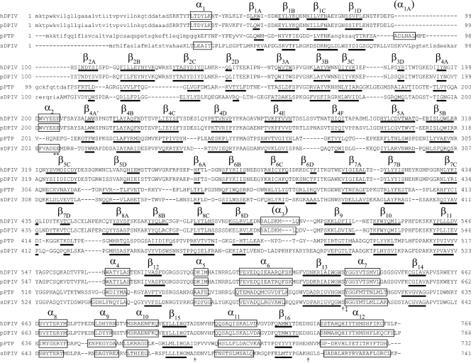
FIG. 2.
Structure-based sequence alignment and associated secondary structures of human, porcine, and Stenotrophomonas DPIVs. The primary structures are aligned on the basis of a three-dimensional structure, using DaLiLite (44). hDPIV, pDPIV, and sDPIV indicate human, porcine, and Stenotrophomonas DPIVs, respectively. Capital and lowercase letters show the parts where the structure is known and unknown, respectively. The secondary structural elements are indicated by boxes labeled α for α-helices and by bold lines labeled β for β-strands. The catalytic triad and residues comprising the hydrophobic pocket are indicated by daggers and double daggers, respectively. Asterisks indicate two glutamate residues recognizing the substrate N-terminal amino group.

