
Fig. 1.
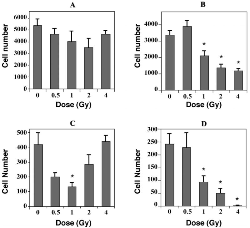
The effects of 56Fe-particle radiation on newly born cells in the dentate subgranular zone of mice. One month after irradiation mice received a single daily injection of 5-bromo-2′deoxyuridine (BrdU) for 7 consecutive days. Three weeks later, tissues were collected and immunohistochemistry and confocal microscopic analyses were done to quantify the numbers of cells co-labeled with BrdU and cell-specific antibodies. Cell numbers represented an estimate of the total number of positively labeled cells in the subgranular zone in both hemispheres. Values for irradiated mice were compared to those for nonirradiated controls. Panel A: Total numbers of BrdU-positive cells decreased slightly over the dose range of 0–2 Gy; the increase after 4 Gy largely represents newly born activated microglia (see Fig. 2). Panel B: Newly born neurons, which were labeled with an antibody against NeuN, showed significant decreases at all doses greater than 0.5 Gy. Panel C: Newly born astrocytes, labeled with an antibody against GFAP, showed substantial decreases after 0.5 and 1.0 Gy. The relative increases seen from 2–4 Gy likely represent a reactive gliosis but could also involve, in part, a regenerative response in GFAP-producing stem cells. Panel D: Newly born oligodendrocytes, which were labeled with an antibody against NG2, showed significant decreases after doses greater than 0.5 Gy. Each bar represents the mean for five or six mice; error bars are standard errors of the means. *P < 0.05 after adjustment for multiple comparisons.