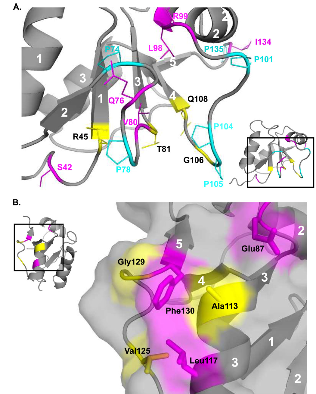
Figure 6.
Two views of slow motions in Pol λ BRCT domain. Residues that demonstrated Rex from CPMG experiments are colored magenta, and residues with multiple peaks in the 15N-1H HSQC are colored yellow. Proline residues are colored cyan. (A). Numerous residues in the vicinity of prolines that display either Rex or slow exchange cross-peaks. (B). A patch of surface-exposed residues that are mostly hydrophobic, and also show either Rex or slow exchange cross-peaks.

