Figure 10.

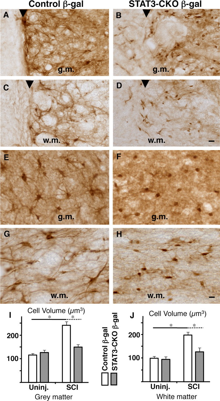
Failure of astrocyte hypertrophy and scar formation after SCI in GFAP-STAT3-CKO mice. A–H, Horizontal sections of spinal cord stained by brightfield immunohistochemistry for the reporter protein β-gal 14 d after moderate crush SCI in nontransgenic control (A, C, E, G) and GFAP-STAT3-CKO (B, D, F, H) mice. A–D show tissue directly along the wound margin. E-H show tissue adjacent to the lesion In control mice, SCI induces a pronounced hypertrophy of β-gal-positive astrocytes with formation of an organized scar that precisely demarcates the lesion boundary (arrowheads) in both gray (A, E) and white (C, G) matter. In contrast, in STAT3-CKO mice, astrocytes fail to hypertrophy after SCI and there is no orderly formation of a glial scar that demarcates the lesion boundary (arrowheads) in either gray (g.m.) (B, F) or white (D, H) matter (w.m.). I, J, Graphs showing the mean cell volume of β-gal stained astrocytes in control and GFAP-STAT3-CKO mice, uninjured (Uninj.), and 14 d after SCI. Stereological measurements of cell volume were performed in a 200 μm boundary zone traced around the entire lesion in both gray and white matter. Astrocytes in both gray (I) and white (J) matter undergo significant hypertrophy after SCI in control mice, but not in STAT3-CKO mice. *p < 0.05 ANOVA with post hoc pairwise analysis; n = 4 or more per group. Scale bars: (in D) A–D, 30 μm; (in H) E–H, 10 μm. Error bars indicate SEM.
