
Figure 2.
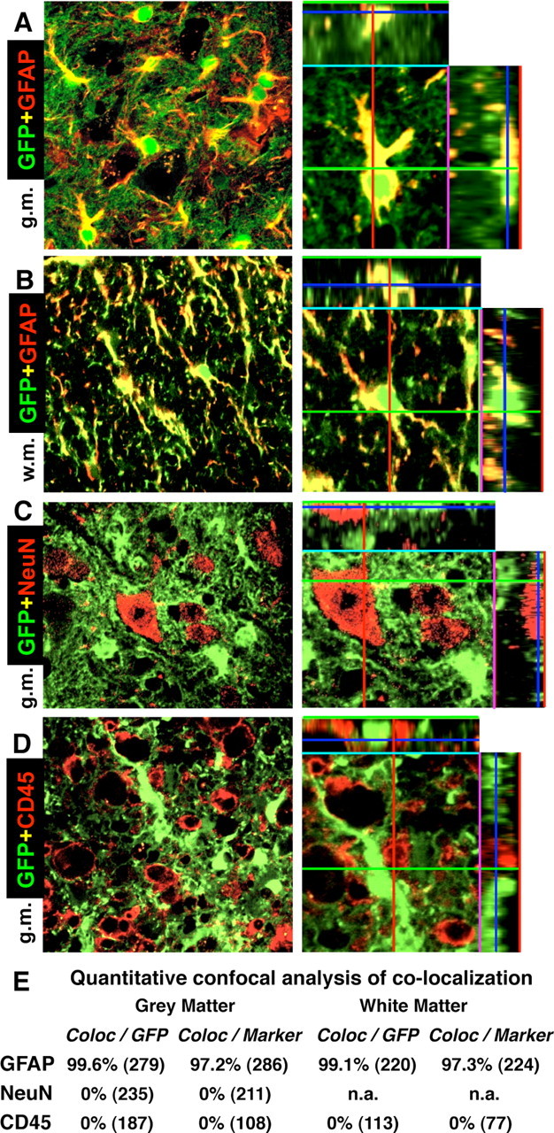
A–D, Specificity of Cre-mediated loxP recombination in astrocytes of 73.12 GFAP-Cre reporter mice at 7 d after SCI in lumbar spinal cord at the immediate margin (A–C) or in the core (D) of the lesion. Survey and detail (ortho) confocal microscopy of the reporter protein GFP combined with immunofluorescence of marker proteins for various cell types is shown. A, B, All cells that express the reporter GFP, also express the astrocyte marker GFAP, and vice versa, in both gray (A) and white (B) matter. Note that the reporter GFP is present in the cell body and throughout many fine distal astrocyte processes, whereas GFAP is present primarily in large proximal processes. C, No cells that express the neuronal marker NeuN also express GFP. D, No cells that express the microglial and white blood cell marker CD45 also express GFP. E, Quantitative confocal analysis of colocalization of reporter GFP and cell type markers. The total numbers of cells evaluated from n = 4 mice are shown in parentheses. Values are expressed as the percentage of cells that exhibit colocalization. g.m., Gray matter; w.m., white matter.