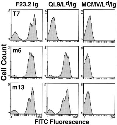
Figure 1.
Flow cytometric analysis of yeast cells that express wild-type and mutant 2C TCR on their surface. Yeast cells displaying wild-type (T7) and mutant (m6 and m13) scTCR were stained with anti-Vβ8 antibody F23.2 (120 nM), the specific alloantigenic peptide-MHC, QL9/Ld/Ig (40 nM), or a null peptide MCMV/Ld/Ig (40 nM). The peptides used in this study were QL9 (QLSPFPFDL), MCMV (YPHFMPTNL), and p2Ca (LSPFPFDL). Binding was detected by FITC-conjugated goat anti-mouse IgG F(ab′)2 and analyzed by flow cytometry. The negative population (e.g., seen with F23.2 staining) has been observed for all yeast-displayed proteins and is thought to be caused by cells at a stage of growth or induction that are incapable of expressing surface fusion protein (16, 17, 27).

