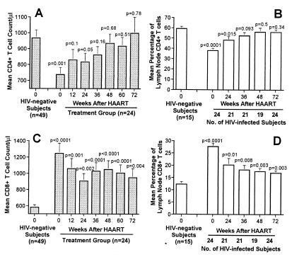
Figure 2.
Changes in CD4 and CD8 T cell counts in blood and percentage in lymph nodes following HAART. (A) Mean CD4 T cell counts after HAART. (B) Mean CD4 T cell percentage in lymph nodes after HAART. (C) Mean CD8 T cell counts after HAART. (D) Mean CD8 T cell percentage in lymph nodes after HAART. Determination of CD4 and CD8 T cell percentages was performed on mononuclear cell populations obtained by excisional lymph node biopsies at weeks 0 and 72 and by ultrasound-guided lymph node aspirates at weeks, 24, 36, and 48. The group of 49 HIV-negative subjects was matched for age and gender with the treatment group. In HIV-negative subjects, determinations of CD4 and CD8 T cell percentages were performed on mononuclear cell populations obtained by excisional lymph node biopsies. Differences in the means between HIV-negative and HIV-positive subjects were compared by the F-test and Student's t test.

