Figure 2.
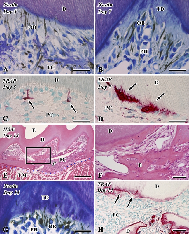
Nestin immunoreactivity (A,B,G), tartrate-resistant acid phosphatase (TRAP) reactions (C,D,H), and H&E staining (E,F) in sections of the transplanted teeth at 5 (A,C), 7 (B,D), and 14 (E–H) days after operation (B, bone-like tissue; D, dentin; E, enamel space; LM, lingual muscle; PC, pulp chamber; PH, pulp horn; TD, tubular dentin). (A,B,G) Newly differentiated odontoblast-like cells (OB) in the pulp horn show intense immunoreactivity for nestin in their cytoplasm. (C,D) Intense TRAP-positive cells appear in the pulp chamber, where they are occasionally situated at the pulp–dentin border and elongate their cellular processes into the dentinal tubules (arrows). (E) Bone-like tissue is formed in the pulp chamber. (F) Higher magnification of boxed area labeled by F in E. The distinction between dentin and bone-like tissue is determined by the absence of dentinal tubules and the existence of cell inclusion in the case of bone-like tissue. (H) TRAP-positive cells remain around the bone matrix. Note the TRAP-positive reactions along the pulp–dentin border (arrows). Blue dotted line shows the boundary between the pulp chamber and surrounding lingual muscle tissue. Bars: E = 250 μm; F,H = 50 μm; A–D,G = 25 μm.

