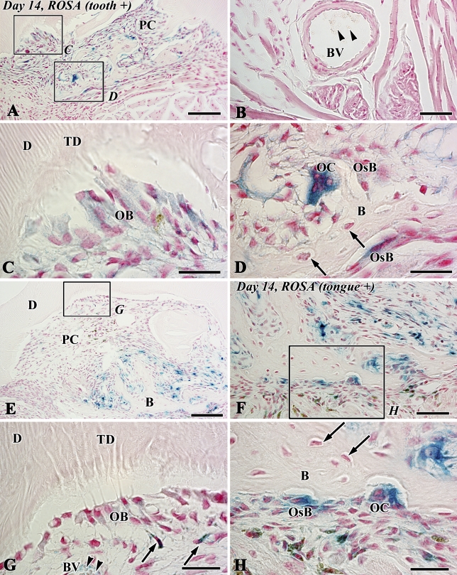
Figure 6.
X-gal staining in sections of the transplants at 14 days after allograft of the tooth of LacZ transgenic ROSA26 mice into the sublingual region of wild-type mice (A–D) and vice versa (E–H) (B, bone-like tissue; D, dentin; PC, pulp chamber; TD, tubular dentin). (A,C,D) Newly differentiated odontoblast-like cells (OB) beneath the dentin matrix contain granular depositions with the blue color. Osteoblast-like cells (OsB) also show blue color in their cytoplasm in addition to the intense stainability in the polynuclear giant cells, osteoclast-like cells (OC). Note osteocytes with negative reactions (arrows) in the bone matrix. (B) A blood vessel in the lingual tissue contains red blood cells with negative reactions (arrowheads). (C,D) Higher magnification of boxed areas labeled by C and D in A, respectively. (E–H) Blue-stained cells are recognized in osteoclast-like cells (OC), osteoblast-like cells (OsB), mesenchymal cells (arrows in G), and the red blood cells (arrowheads) in blood vessels (BV) in contrast with odontoblast-like cells (OB) and osteocytes (arrows in H) with negative reactions. (G,H) Higher magnification of boxed areas labeled by G and H in E and F, respectively. Bars: A,E = 100 μm; B,F = 50 μm; C,D,G,H = 25 μm.

