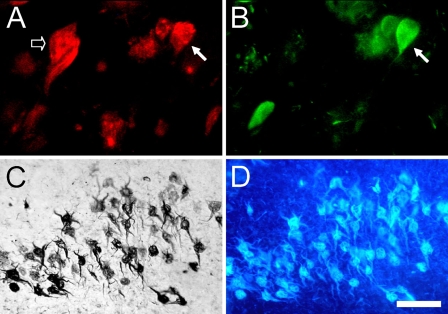
Figure 7.
Dual immunofluorescence for 5-LOX (Santa Cruz-N; red) and tau (green) in the hippocampus (A,B) from an AD patient. Solid arrows point to the same cell that is positive for 5-LOX and tau. Some of the 5-LOX-ir cells (A, empty arrow) do not contain tau accumulation. Combined 5-LOX IHC (C, DAB/nickel) and X-34 histochemistry (D, blue fluorescence) in the same entorhinal cortex tissue section show that, in advanced AD, all of the 5-LOX-ir lamina II neurons also contain pathological β-pleated sheet conformed protein aggregates. Bar: A,B = 25 μm; C,D = 50 μm.

