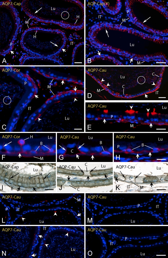
Figure 4.
Light micrographs of caput (Cap) (A,B,I), corpus (Cor) (C,F), and cauda (Cau) (D,E,G,H,J–O) regions of epididymis from adult (A) (C–O) and young (Y) (B) rats in zinc-fixed (A–H,L–O) and Ste. Marie–fixed (I–K) tissues immunostained for AQP 7. Three different antibodies to rat AQP 7 were used including an R-101 antibody from Santa Cruz (A–K) and synthetic peptide antibodies to short sequences at the N-terminal (L–N) and C-terminal (O) ends of AQP 7. In M, the N-terminal antibody was preincubated with its specific blocking peptide, whereas in N, the same antibody was preincubated with an unrelated peptide of similar length. The site of localization for AQP 7 in principal cells varies by antibody, region, and cell type. B, basal cells; C, clear cells; H, halo cells; IT, intertubular space; Lu, lumen; M, myoid cells; P, principal cells; arrowheads, apical membrane reaction; long arrows, lateral membrane reaction; short arrows, basal membrane reaction; circle, reactive sperm. Bars: A,B,D = 40 μm; C,E,J–O = 20 μm; F–H = 10 μm; I = 15 μm.

