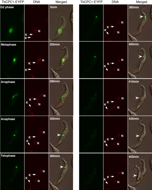
Figure 1. Translocalization of TbCPC1-EYFP in live cell imaged by video fluorescence microscopy.
Procyclic cell stably expressing the enhanced yellow fluorescent protein (EYFP) tagged TbCPC1 was imaged during mitosis-cytokinesis. DNA was stained with Hoechst DNA dye, and the fluorescence and phase images were merged. Selected images at various time intervals are shown to illustrate the trans-localization of TbCPC1-EYFP (arrowheads) from nucleus to spindle midzone and then to dorsal side toward the anterior end and finally to the posterior end of the cell. Nucleus (N) and kinetoplast DNA (K) are indicated. The complete image sequence is available as a video in the Supplementary file Movie S1.

