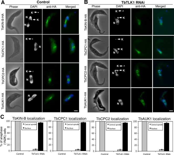
Figure 3. Effects of TbTLK1 knockdown on subcellular localizations of TbKIN-B, TbCPC1, TbCPC2 and TbAUK1.
(A). TbKIN-B-3HA, TbCPC1-3HA, TbCPC2-3HA and TbAUK1-3HA expressed to the endogenous levels in un-induced RNAi cells were detected with FITC-conjugated anti-HA mAb. (B). Localization of the 3HA-tagged TbKIN-B, TbCPC1, TbCPC2 and TbAUK1-3HA after a 24 hr RNAi of TbTLK1. All stained cells were of the 1N2K type (N, nucleus; K, kinetoplast) in the anaphase stage of cell cycle. (C). Percentages of 1N2K cells with TbKIN-B-3HA, TbCPC1-3HA, TbCPC2-3HA and TbAUK1-3HA localized to the central spindle. Data are presented as the mean percent ±S.D. of ∼300 cells counted from three independent experiments. Bars: 2 µm.

