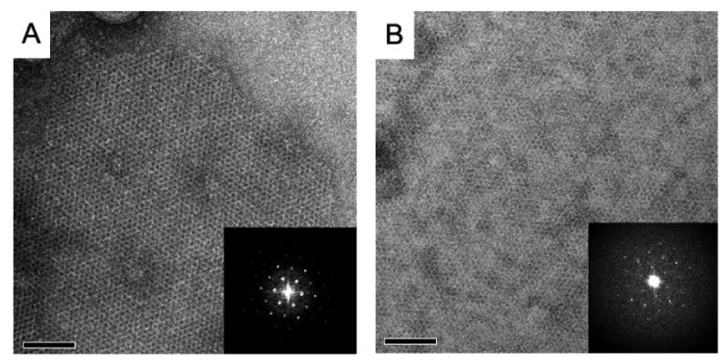
Fig. 2.

Two types of 2D crystals imaged in negative stain. (A) The 65-kDa Cry4Ba toxin crystal grown in DMPC on the air glow discharged carbon surface, and (B) the toxin crystal in DMPC attached on the hydrophobic carbon surface. Crystals were up to 1×1 μm in size. Insets show Fourier transforms of these crystalline areas.
