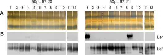
Figure 4. LPS profiles of H. pylori isolates after large bottleneck in vitro passages (50pL).
Silver staining (A) and Immunoblot analysis, detecting Lex and Ley (B), of extracted LPS from twelve single-colony isolates per strain, obtained after 50 in vitro passages of bacterial sweeps on agar medium, reveals minor differences in the LPS molecules.

