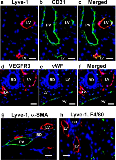
Figure 1. Lymphatic endothelial cells are readily distinguished from vascular endothelial cells.
The portal triad region of mouse liver was analyzed for the expression of lymphatic (Lyve-1, VEGFR-3) and vascular (CD31, vWF) endothelial cell markers, a macrophage marker (F4/80) and a pericyte/smooth muscle marker (α-SMA). (a) Lymphatic endothelial cells are Lyve-1+ (red). (b) Both portal veins and lymphatic vessels express CD31 (green). (c) Merged image of a and b. Double positive CD31+, Lyve-1+ cells are yellow. d) VEGFR3+ lymphatic endothelial cells (red), do not express (e) the blood vessel endothelial marker vWF (green). (f) Merged image of d and e. (g) Lyve-1+ lymphatic vessels (red) do not express the pericyte marker α-SMA (green) or (h) the macrophage marker F4/80 (green). (DAPI stained nuclei are blue. PV = portal vein, LV = lymphatic vessel, BD = bile duct; scale bars: 20 µm).

