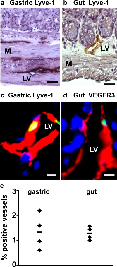
Figure 3. HSC-derived lymphatic endothelial cells are detected in gastrointestinal tissues.
(a & b) Bright field images of (a) gastric and (b) gut tissue containing Lyve-1+ (brown, DAB-stained) lymphatic vessels. Sections are counterstained with hematoxylin (purple). (c) Merged image of a donor-derived (green), Lyve-1+ (red) lymphatic endothelial cell in the stomach. (d) Merged image of donor-derived (green), VEGFR-3+ (red) lymphatic endothelial cell in the gut. DAPI stained nuclei are blue. (e) Frequency of lymphatic vessels in the stomach and gut containing donor-derived endothelial cells after HSC transplantation. Each symbol represents an individual HSC recipient. The horizontal line indicates the average for each group. (LV = Lymphatic Vessel, M = Muscle. Scale bars: a: 50 µm; b: 20 µm; c & d: 5 µm).

