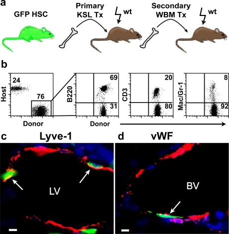
Figure 4. Lymphatic and vascular endothelial potential of self-renewing of HSCs.
(a) Secondary transplantation scheme. (b) Flow cytometry analysis of multi-lineage hematopoietic cells in the peripheral blood of secondary recipients. Donor-derived (GFP+) B220 (B-cells), CD3 (T-cells), Mac1/Gr-1(myeloid cells) are shown. (c) Merged image of donor-derived (green), Lyve-1+ (red), CD45− (absence of magenta) lymphatic endothelial cells (arrows). (d) Merged image of donor-derived (green), vWF+ (red), CD45- (absence of magenta) vascular endothelial cell (arrow). (DAPI stained nuclei are blue in c,d. LV = lymphatic vessel, BV = blood vessel; scale bars: 5 µm).

