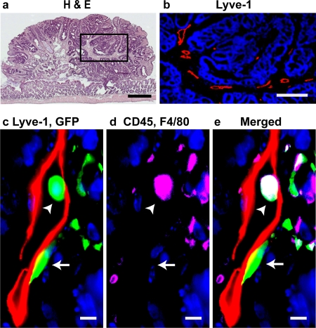
Figure 5. HSC-derived lymphatic endothelial cells incorporate into tumor lymphatics.
Following transplantation of HSCs into Min mice, spontaneously developing intestinal tumors were examined for donor-derived (GFP+), Lyve-1+, CD45−, F4/80− lymphatic endothelial cells. (a) Hematoxylin and eosin (H&E) staining of a tumor in a Min mouse. (b) High magnification of the boxed area in panel a demonstrating Lyve-1+ (red) lymphatic vessels within the tumor. (c) Merged image of a donor-derived (green), Lyve-1+ (red) lymphatic endothelial cell (arrow) and a donor-derived hematopoietic cell (arrow head). (d) The donor-derived hematopoietic cell expresses CD45 and/or F4/80 (magenta), whereas the donor-derived LEC does not. (e) Merged image of c and d (green+ blue+ magenta = white). (DAPI stained nuclei are blue in b–e. Scale bars: a: 500 µm; b: 200 µm, c–e: 5 µm)

