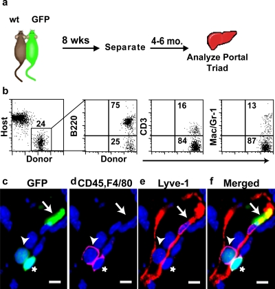
Figure 6. Parabiosis reveals the presence of functional circulating lymphatic progenitors in the absence of acute tissue injury.
(a) Non-irradiated wild type mice were surgically joined (parabiosed) with GFP partners. After 12 weeks, mice were separated for between 4–6 months then lymphatic vessels were analyzed. (b) Separated, individual mice displayed stable, donor-derived multi-lineage hematopoietic reconstitution of the peripheral blood. (c–f) Analysis of donor-derived cells in and near a lymphatic vessel. A donor-derived LEC (arrow), a donor-derived hematopoieitc cell within the lymphatic vessel (arrow head), and a donor-derived hematopoietic cell adjacent to the lymphatic vessel (asterisk) are shown. DAPI stained nuclei are blue. Scale bars: 5 µm. (c) Donor-derived, GFP+ cells (green). (d) CD45 and F4/80 distinguishes hematopoietic (magenta, arrowhead and asterisk) from non-hematopoietic (arrow) donor-derived cells. (e) Lyve-1+ (red) reveals the presence of a donor-derived LEC (arrow). (f) Merged image of c–e.

