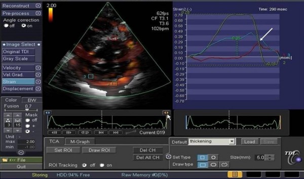
Figure 5.

Example of a strain curve in a patient with anterior myocardial infarction. The green curve sampled from the normocontractile posterolateral wall shows a pronounced systolic deformation; in the red curve derived from the ischemic interventricular septum only minimal systolic myocardial deformation followed by post-systolic shortening can be seen (see arrow).
